Actin, alpha skeletal muscle
Protein-coding gene in the species Homo sapiens
| ACTA1 | |||||||||||||||||||||||||||||||||||||||||||||||||||
|---|---|---|---|---|---|---|---|---|---|---|---|---|---|---|---|---|---|---|---|---|---|---|---|---|---|---|---|---|---|---|---|---|---|---|---|---|---|---|---|---|---|---|---|---|---|---|---|---|---|---|---|
 | |||||||||||||||||||||||||||||||||||||||||||||||||||
| Identifiers | |||||||||||||||||||||||||||||||||||||||||||||||||||
| Aliases | ACTA1, ACTA, ASMA, CFTD, CFTD1, CFTDM, MPFD, NEM1, NEM2, NEM3, Actin, alpha 1, SHPM, actin, alpha 1, skeletal muscle, actin alpha 1, skeletal muscle | ||||||||||||||||||||||||||||||||||||||||||||||||||
| External IDs | OMIM: 102610; MGI: 87902; HomoloGene: 121702; GeneCards: ACTA1; OMA:ACTA1 - orthologs | ||||||||||||||||||||||||||||||||||||||||||||||||||
| |||||||||||||||||||||||||||||||||||||||||||||||||||
| |||||||||||||||||||||||||||||||||||||||||||||||||||
| |||||||||||||||||||||||||||||||||||||||||||||||||||
| |||||||||||||||||||||||||||||||||||||||||||||||||||
| |||||||||||||||||||||||||||||||||||||||||||||||||||
| Wikidata | |||||||||||||||||||||||||||||||||||||||||||||||||||
| |||||||||||||||||||||||||||||||||||||||||||||||||||


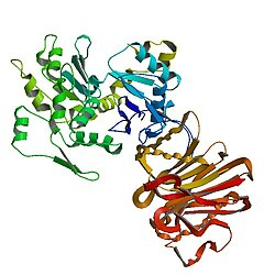
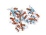
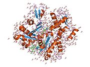
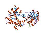
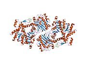
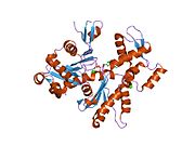
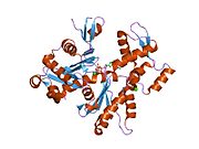
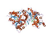
Actin, alpha skeletal muscle is a protein that in humans is encoded by the ACTA1 gene.[5][6]
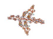
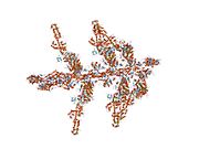
Actin alpha 1 which is expressed in skeletal muscle is one of six different actin isoforms which have been identified. Actins are highly conserved proteins that are involved in cell motility, structure and integrity. Alpha actins are a major constituent of the contractile apparatus.[7]
Skeletal actin gene expression
Skeletal alpha actin expression is induced by stimuli and conditions known to cause muscle formation.[8] Such conditions result in fusion of committed cells (satellite cells) into myotubes, to form muscle fibers. Skeletal actin itself, when expressed, causes expression of several other "myogenic genes", which are essential to muscle formation.[9] One key transcription factor that activates skeletal actin gene expression is Serum Response Factor ("SRF"), a protein that binds to specific sites on the promoter DNA of the actin gene.[10] SRF may bring a number of other proteins to the promoter of skeletal actin, such as androgen receptor, and thereby contribute to induction of skeletal actin gene expression by androgenic (often termed "anabolic") steroids.[11]
Interactions
Actin, alpha 1 has been shown to interact with TMSB4X,[12][13] MIB2[14] and PRKCE.[15]
Clinical significance
Mutations in the ACTA1 gene are known to cause the following conditions:[16]
- Nemaline myopathy 3 (NEM3);
- Myopathy, actin, congenital, with excess of thin myofilaments (MPCETM);
- Myopathy, congenital, with fiber-type disproportion (CFTD);
- Myopathy, scapulohumeroperoneal (SHPM).
See also
References
- ^ a b c GRCh38: Ensembl release 89: ENSG00000143632 – Ensembl, May 2017
- ^ a b c GRCm38: Ensembl release 89: ENSMUSG00000031972 – Ensembl, May 2017
- ^ "Human PubMed Reference:". National Center for Biotechnology Information, U.S. National Library of Medicine.
- ^ "Mouse PubMed Reference:". National Center for Biotechnology Information, U.S. National Library of Medicine.
- ^ Mogensen J, Kruse TA, Børglum AD (March 1999). "Assignment of the human skeletal muscle [FC12]a-actin gene (ACTA1) to chromosome 1q42.13→q42.2 by radiation hybrid mapping". Cytogenetics and Cell Genetics. 83 (3–4): 224–5. doi:10.1159/000015184. PMID 10072583. S2CID 84202330.
- ^ Gunning P, Ponte P, Okayama H, Engel J, Blau H, Kedes L (May 1983). "Isolation and characterization of full-length cDNA clones for human alpha-, beta-, and gamma-actin mRNAs: skeletal but not cytoplasmic actins have an amino-terminal cysteine that is subsequently removed". Molecular and Cellular Biology. 3 (5): 787–95. doi:10.1128/mcb.3.5.787. PMC 368601. PMID 6865942.
- ^ "Entrez Gene: ACTA1 actin, alpha 1, skeletal muscle".
- ^ Bandman E (December 1992). "Contractile protein isoforms in muscle development". Developmental Biology. 154 (2): 273–83. doi:10.1016/0012-1606(92)90067-Q. PMID 1358730.
- ^ Gunning PW, Ferguson V, Brennan KJ, Hardeman EC (February 2001). "Alpha-skeletal actin induces a subset of muscle genes independently of muscle differentiation and withdrawal from the cell cycle". Journal of Cell Science. 114 (Pt 3): 513–24. doi:10.1242/jcs.114.3.513. PMID 11171321.
- ^ Belaguli NS, Zhou W, Trinh TH, Majesky MW, Schwartz RJ (July 1999). "Dominant negative murine serum response factor: alternative splicing within the activation domain inhibits transactivation of serum response factor binding targets". Molecular and Cellular Biology. 19 (7): 4582–91. doi:10.1128/mcb.19.7.4582. PMC 84256. PMID 10373507.
- ^ Vlahopoulos S, Zimmer WE, Jenster G, Belaguli NS, Balk SP, Brinkmann AO, Lanz RB, Zoumpourlis VC, Schwartz RJ (March 2005). "Recruitment of the androgen receptor via serum response factor facilitates expression of a myogenic gene". The Journal of Biological Chemistry. 280 (9): 7786–92. doi:10.1074/jbc.M413992200. PMID 15623502.
- ^ Ballweber E, Hannappel E, Huff T, Stephan H, Haener M, Taschner N, Stoffler D, Aebi U, Mannherz HG (January 2002). "Polymerisation of chemically cross-linked actin:thymosin beta(4) complex to filamentous actin: alteration in helical parameters and visualisation of thymosin beta(4) binding on F-actin". Journal of Molecular Biology. 315 (4): 613–25. doi:10.1006/jmbi.2001.5281. PMID 11812134.
- ^ Safer D, Sosnick TR, Elzinga M (May 1997). "Thymosin beta 4 binds actin in an extended conformation and contacts both the barbed and pointed ends". Biochemistry. 36 (19): 5806–16. doi:10.1021/bi970185v. PMID 9153421.
- ^ Takeuchi T, Heng HH, Ye CJ, Liang SB, Iwata J, Sonobe H, Ohtsuki Y (October 2003). "Down-regulation of a novel actin-binding molecule, skeletrophin, in malignant melanoma". The American Journal of Pathology. 163 (4): 1395–404. doi:10.1016/S0002-9440(10)63497-9. PMC 1868282. PMID 14507647.
- ^ England K, Ashford D, Kidd D, Rumsby M (June 2002). "PKC epsilon is associated with myosin IIA and actin in fibroblasts". Cellular Signalling. 14 (6): 529–36. doi:10.1016/S0898-6568(01)00277-7. PMID 11897493.
- ^ "UniProt". www.uniprot.org. Retrieved 2023-09-09.
Further reading
- Snásel J, Pichová I (1997). "The cleavage of host cell proteins by HIV-1 protease". Folia Biologica. 42 (5): 227–30. doi:10.1007/BF02818986. PMID 8997639. S2CID 7617882.
- Di Fiore PP, Scita G (October 2002). "Eps8 in the midst of GTPases". The International Journal of Biochemistry & Cell Biology. 34 (10): 1178–83. doi:10.1016/S1357-2725(02)00064-X. PMID 12127568.
- Ogawa H, Shiraki H, Matsuda Y, Nakagawa H (April 1978). "Interaction of adenylosuccinate synthetase with F-actin". European Journal of Biochemistry. 85 (2): 331–7. doi:10.1111/j.1432-1033.1978.tb12243.x. PMID 648524.
- den Hartigh JC, van Bergen en Henegouwen PM, Verkleij AJ, Boonstra J (October 1992). "The EGF receptor is an actin-binding protein". The Journal of Cell Biology. 119 (2): 349–55. doi:10.1083/jcb.119.2.349. PMC 2289650. PMID 1383230.
- Adams LD, Tomasselli AG, Robbins P, Moss B, Heinrikson RL (February 1992). "HIV-1 protease cleaves actin during acute infection of human T-lymphocytes". AIDS Research and Human Retroviruses. 8 (2): 291–5. doi:10.1089/aid.1992.8.291. PMID 1540415.
- Levine BA, Moir AJ, Patchell VB, Perry SV (February 1992). "Binding sites involved in the interaction of actin with the N-terminal region of dystrophin". FEBS Letters. 298 (1): 44–8. doi:10.1016/0014-5793(92)80019-D. PMID 1544421.
- Rijken PJ, Hage WJ, van Bergen en Henegouwen PM, Verkleij AJ, Boonstra J (November 1991). "Epidermal growth factor induces rapid reorganization of the actin microfilament system in human A431 cells". Journal of Cell Science. 100 ( Pt 3) (3): 491–9. doi:10.1242/jcs.100.3.491. PMID 1808202.
- Tomasselli AG, Hui JO, Adams L, Chosay J, Lowery D, Greenberg B, Yem A, Deibel MR, Zürcher-Neely H, Heinrikson RL (August 1991). "Actin, troponin C, Alzheimer amyloid precursor protein and pro-interleukin 1 beta as substrates of the protease from human immunodeficiency virus". The Journal of Biological Chemistry. 266 (22): 14548–53. doi:10.1016/S0021-9258(18)98721-1. PMID 1907279.
- Shoeman RL, Kesselmier C, Mothes E, Höner B, Traub P (January 1991). "Non-viral cellular substrates for human immunodeficiency virus type 1 protease". FEBS Letters. 278 (2): 199–203. doi:10.1016/0014-5793(91)80116-K. PMID 1991513.
- Winder SJ, Walsh MP (June 1990). "Smooth muscle calponin. Inhibition of actomyosin MgATPase and regulation by phosphorylation". The Journal of Biological Chemistry. 265 (17): 10148–55. doi:10.1016/S0021-9258(19)38792-7. PMID 2161834.
- Kabsch W, Mannherz HG, Suck D, Pai EF, Holmes KC (September 1990). "Atomic structure of the actin:DNase I complex". Nature. 347 (6288): 37–44. Bibcode:1990Natur.347...37K. doi:10.1038/347037a0. PMID 2395459. S2CID 925337.
- Takahashi K, Hiwada K, Kokubu T (June 1988). "Vascular smooth muscle calponin. A novel troponin T-like protein". Hypertension. 11 (6 Pt 2): 620–6. doi:10.1161/01.hyp.11.6.620. PMID 2455687.
- Taylor A, Erba HP, Muscat GE, Kedes L (November 1988). "Nucleotide sequence and expression of the human skeletal alpha-actin gene: evolution of functional regulatory domains". Genomics. 3 (4): 323–36. doi:10.1016/0888-7543(88)90123-1. PMID 2907503.
- Shen BW, Josephs R, Steck TL (March 1986). "Ultrastructure of the intact skeleton of the human erythrocyte membrane". The Journal of Cell Biology. 102 (3): 997–1006. doi:10.1083/jcb.102.3.997. PMC 2114132. PMID 2936753.
- Burgess DR, Broschat KO, Hayden JM (January 1987). "Tropomyosin distinguishes between the two actin-binding sites of villin and affects actin-binding properties of other brush border proteins". The Journal of Cell Biology. 104 (1): 29–40. doi:10.1083/jcb.104.1.29. PMC 2117036. PMID 3793760.
- Kedes L, Ng SY, Lin CS, Gunning P, Eddy R, Shows T, Leavitt J (1986). "The human beta-actin multigene family". Transactions of the Association of American Physicians. 98: 42–6. PMID 3842206.
- Hanauer A, Levin M, Heilig R, Daegelen D, Kahn A, Mandel JL (June 1983). "Isolation and characterization of cDNA clones for human skeletal muscle alpha actin". Nucleic Acids Research. 11 (11): 3503–16. doi:10.1093/nar/11.11.3503. PMC 325982. PMID 6190133.
- Bretscher A, Weber K (July 1980). "Villin is a major protein of the microvillus cytoskeleton which binds both G and F actin in a calcium-dependent manner". Cell. 20 (3): 839–47. doi:10.1016/0092-8674(80)90330-X. PMID 6893424. S2CID 568395.
External links
- GeneReviews/NCBI/NIH/UW entry on Nemaline Myopathy
- Human ACTA1 genome location and ACTA1 gene details page in the UCSC Genome Browser.
- v
- t
- e
PDB gallery
-
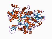
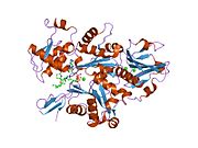
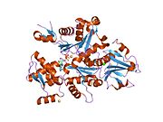
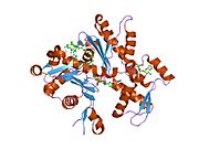
 1atn: Atomic structure of the actin:DNASE I complex
 -
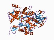
 1c0g: CRYSTAL STRUCTURE OF 1:1 COMPLEX BETWEEN GELSOLIN SEGMENT 1 AND A DICTYOSTELIUM/TETRAHYMENA CHIMERA ACTIN (MUTANT 228: Q228K/T229A/A230Y/E360H)
 -
 1d4x: Crystal Structure of Caenorhabditis Elegans Mg-ATP Actin Complexed with Human Gelsolin Segment 1 at 1.75 A resolution.
 -
 1eqy: COMPLEX BETWEEN RABBIT MUSCLE ALPHA-ACTIN: HUMAN GELSOLIN DOMAIN 1
 -
 1esv: COMPLEX BETWEEN LATRUNCULIN A:RABBIT MUSCLE ALPHA ACTIN:HUMAN GELSOLIN DOMAIN 1
 -
 1h1v: GELSOLIN G4-G6/ACTIN COMPLEX
 -
 1hlu: STRUCTURE OF BOVINE BETA-ACTIN-PROFILIN COMPLEX WITH ACTIN BOUND ATP PHOSPHATES SOLVENT ACCESSIBLE
 -
 1ijj: THE X-RAY CRYSTAL STRUCTURE OF THE COMPLEX BETWEEN RABBIT SKELETAL MUSCLE ACTIN AND LATRUNCULIN A AT 2.85 A RESOLUTION
 -
 1j6z: UNCOMPLEXED ACTIN
 -
 1kxp: CRYSTAL STRUCTURE OF HUMAN VITAMIN D-BINDING PROTEIN IN COMPLEX WITH SKELETAL ACTIN
 -
 1lcu: Polylysine Induces an Antiparallel Actin Dimer that Nucleates Filament Assembly: Crystal Structure at 3.5 A Resolution
 -
 1lot: CRYSTAL STRUCTURE OF THE COMPLEX OF ACTIN WITH VITAMIN D-BINDING PROTEIN
 -
 1m8q: Molecular Models of Averaged Rigor Crossbridges from Tomograms of Insect Flight Muscle
 -
 1ma9: Crystal structure of the complex of human vitamin D binding protein and rabbit muscle actin
 -
 1mdu: Crystal structure of the chicken actin trimer complexed with human gelsolin segment 1 (GS-1)
 -
 1mvw: MOLECULAR MODELS OF AVERAGED RIGOR CROSSBRIDGES FROM TOMOGRAMS OF INSECT FLIGHT MUSCLE
 -
 1nlv: Crystal Structure Of Dictyostelium Discoideum Actin Complexed With Ca ATP And Human Gelsolin Segment 1
 -
 1nm1: Crystal Structure of D. Dicsoideum Actin Complexed With Gelsolin Segment 1 and Mg ATP at 1.8 A Resolution
 -
 1nmd: Crystal Structure of D. Discoideum Actin-Gelsolin Segment 1 Complex Crystallized In Presence Of Lithium ATP
 -
 1nwk: CRYSTAL STRUCTURE OF MONOMERIC ACTIN IN THE ATP STATE
 -
 1o18: MOLECULAR MODELS OF AVERAGED RIGOR CROSSBRIDGES FROM TOMOGRAMS OF INSECT FLIGHT MUSCLE
 -
 1o19: MOLECULAR MODELS OF AVERAGED RIGOR CROSSBRIDGES FROM TOMOGRAMS OF INSECT FLIGHT MUSCLE
 -
 1o1a: MOLECULAR MODELS OF AVERAGED RIGOR CROSSBRIDGES FROM TOMOGRAMS OF INSECT FLIGHT MUSCLE
 -
 1o1b: MOLECULAR MODELS OF AVERAGED RIGOR CROSSBRIDGES FROM TOMOGRAMS OF INSECT FLIGHT MUSCLE
 -
 1o1c: MOLECULAR MODELS OF AVERAGED RIGOR CROSSBRIDGES FROM TOMOGRAMS OF INSECT FLIGHT MUSCLE
 -
 1o1d: MOLECULAR MODELS OF AVERAGED RIGOR CROSSBRIDGES FROM TOMOGRAMS OF INSECT FLIGHT MUSCLE
 -
 1o1e: MOLECULAR MODELS OF AVERAGED RIGOR CROSSBRIDGES FROM TOMOGRAMS OF INSECT FLIGHT MUSCLE
 -
 1o1f: MOLECULAR MODELS OF AVERAGED RIGOR CROSSBRIDGES FROM TOMOGRAMS OF INSECT FLIGHT MUSCLE
 -
 1o1g: MOLECULAR MODELS OF AVERAGED RIGOR CROSSBRIDGES FROM TOMOGRAMS OF INSECT FLIGHT MUSCLE
 -
 1p8z: Complex Between Rabbit Muscle alpha-Actin: Human Gelsolin Residues Val26-Glu156
 -
 1qz5: Structure of rabbit actin in complex with kabiramide C
 -
 1qz6: Structure of rabbit actin in complex with jaspisamide A
 -
 1rdw: Actin Crystal Dynamics: Structural Implications for F-actin Nucleation, Polymerization and Branching Mediated by the Anti-parallel Dimer
 -
 1rfq: Actin Crystal Dynamics: Structural Implications for F-actin Nucleation, Polymerization and Branching Mediated by the Anti-parallel Dimer
 -
 1rgi: Crystal structure of gelsolin domains G1-G3 bound to actin
 -
 1s22: Absolute Stereochemistry of Ulapualide A
 -
 1sqk: CRYSTAL STRUCTURE OF CIBOULOT IN COMPLEX WITH SKELETAL ACTIN
 -
 1t44: Structural basis of actin sequestration by thymosin-B4: Implications for arp2/3 activation
 -
 1wua: The structure of Aplyronine A-actin complex
 -
 1y64: Bni1p Formin Homology 2 Domain complexed with ATP-actin
 -
 1yxq: Crystal structure of actin in complex with swinholide A
 -
 2a3z: Ternary complex of the WH2 domain of WASP with Actin-DNAse I
 -
 2a40: Ternary complex of the WH2 domain of WAVE with Actin-DNAse I
 -
 2a41: Ternary complex of the WH2 Domain of WIP with Actin-DNAse I
 -
 2a42: Actin-DNAse I Complex
 -
 2a5x: Crystal Structure of a Cross-linked Actin Dimer
 -
 2asm: Structure of Rabbit Actin In Complex With Reidispongiolide A
 -
 2aso: Structure of Rabbit Actin In Complex With Sphinxolide B
 -
 2asp: Structure of Rabbit Actin In Complex With Reidispongiolide C
 -
 2btf: THE STRUCTURE OF CRYSTALLINE PROFILIN-BETA-ACTIN
 -
 2d1k: Ternary complex of the WH2 domain of mim with actin-dnase I
 -
 2ff3: Crystal structure of Gelsolin domain 1:N-wasp V2 motif hybrid in complex with actin
 -
 2ff6: Crystal structure of Gelsolin domain 1:ciboulot domain 2 hybrid in complex with actin
 -
 2fxu: X-ray Structure of Bistramide A- Actin Complex at 1.35 A resolution.
 -
 2gwj: SpvB ADP-ribosylated actin: hexagonal crystal form
 -
 2gwk: SpvB ADP-ribosylated actin: orthorhombic crystal form
 -
 2hf3: Crystal structure of monomeric Actin in the ADP bound state
 -
 2hf4: Crystal structure of Monomeric Actin in its ATP-bound state
 -
 2hmp: Uncomplexed actin cleaved with protease ECP32
 -
 2oan: Structure of oxidized beta-actin
 -
 2q1n: Actin Dimer Cross-linked Between Residues 41 and 374
 -
 2q31: Actin Dimer Cross-linked Between Residues 41 and 374 and proteolytically cleaved by subtilisin between residues 47 and 48.
 -
 2q36: Actin Dimer Cross-linked between Residues 191 and 374 and complexed with Kabiramide C


