ICAM-1

ICAM1
Structures disponibles
PDBRecherche d'orthologue: PDBe RCSB
Identifiants PDB

1D3E, 1D3I, 1D3L, 1IAM, 1IC1, 1MQ8, 1P53, 1Z7Z, 2OZ4, 3TCX

Identifiants
AliasesICAM1
IDs externesOMIM: 147840 MGI: 96392 HomoloGene: 168 GeneCards: ICAM1
Position du gène (Homme)
Chromosome 19 humain
Chr.Chromosome 19 humain[1]
Chromosome 19 humain
Localisation génomique pour ICAM1
Locus19p13.2Début10,271,093 bp[1]
Fin10,286,615 bp[1]
Position du gène (Souris)
Chromosome 9 (souris)
Chr.Chromosome 9 (souris)[2]
Chromosome 9 (souris)
Localisation génomique pour ICAM1
Locus9 A3|9 7.69 cMDébut20,927,281 bp[2]
Fin20,940,113 bp[2]
Expression génétique
Bgee
HumainSouris (orthologue)
Fortement exprimé dans
  • veine cave

  • upper lobe of left lung

  • poumon droit

  • vésicule biliaire

  • lobe inférieur du poumon

  • rate

  • monocyte

  • ganglion lymphatique

  • left uterine tube

  • appendice iléo-cæcal
Fortement exprimé dans
  • lobe pulmonaire droit

  • poumon gauche

  • left lung lobe

  • rate

  • articulation talo-crurale

  • thymus

  • valve aortique

  • Vésicule vitelline

  • calvaria

  • corpuscule carotidien
Plus de données d'expression de référence
BioGPS


Plus de données d'expression de référence
Gene Ontology
Fonction moléculaire
  • virus receptor activity
  • liaison de complexe macromoléculaire
  • integrin binding
  • liaison protéique
  • transmembrane signaling receptor activity
  • signaling receptor activity
Composant cellulaire
  • integral component of membrane
  • membrane
  • focal adhesion
  • membrane plasmique
  • integral component of plasma membrane
  • surface cellulaire
  • Synapse immunologique
  • radeau lipidique
  • exosome
  • external side of plasma membrane
  • milieu extracellulaire
  • matrice extracellulaire
  • collagen-containing extracellular matrix
Processus biologique
  • leukocyte cell-cell adhesion
  • response to ionizing radiation
  • establishment of endothelial barrier
  • negative regulation of endothelial cell apoptotic process
  • cellular response to organic substance
  • heterophilic cell-cell adhesion via plasma membrane cell adhesion molecules
  • response to amino acid
  • response to hypoxia
  • positive regulation of actin filament polymerization
  • response to organic cyclic compound
  • T cell antigen processing and presentation
  • establishment of Sertoli cell barrier
  • response to sulfur dioxide
  • response to copper ion
  • interferon-gamma-mediated signaling pathway
  • positive regulation of nitric oxide biosynthetic process
  • response to amphetamine
  • cellular response to tumor necrosis factor
  • regulation of leukocyte mediated cytotoxicity
  • T cell activation via T cell receptor contact with antigen bound to MHC molecule on antigen presenting cell
  • extracellular matrix organization
  • acute inflammatory response to antigenic stimulus
  • sensory perception of sound
  • cellular response to alkaloid
  • response to gonadotropin
  • positive regulation of cellular extravasation
  • positive regulation of GTPase activity
  • response to lipopolysaccharide
  • regulation of cell adhesion
  • adhésion cellulaire
  • cell adhesion mediated by integrin
  • positive regulation of NF-kappaB transcription factor activity
  • negative regulation of extrinsic apoptotic signaling pathway via death domain receptors
  • positive regulation of vasoconstriction
  • cellular response to interleukin-1
  • cellular response to nutrient levels
  • negative regulation of calcium ion transport
  • regulation of cell shape
  • membrane to membrane docking
  • positive regulation of peptidyl-tyrosine phosphorylation
  • ovarian follicle development
  • regulation of immune response
  • positive regulation of ERK1 and ERK2 cascade
  • regulation of ruffle assembly
  • pénétration virale
  • response to ethanol
  • viral process
  • cellular response to lipopolysaccharide
  • cellular response to hypoxia
  • leukocyte migration
  • cellular response to glucose stimulus
  • response to insulin
  • cellular response to interferon-gamma
  • cellular response to interleukin-6
  • cellular response to dexamethasone stimulus
  • establishment of endothelial intestinal barrier
  • positive regulation of leukocyte adhesion to vascular endothelial cell
  • cellular response to leukemia inhibitory factor
  • cell-cell adhesion
  • cytokine-mediated signaling pathway
  • T cell extravasation
  • cellular response to amyloid-beta
Sources:Amigo / QuickGO
Orthologues
EspècesHommeSouris
Entrez

3383

15894

Ensembl

ENSG00000090339

ENSMUSG00000037405

UniProt

P05362

P13597

RefSeq (mRNA)

NM_000201

NM_010493

RefSeq (protéine)

NP_000192

NP_034623

Localisation (UCSC)Chr 19: 10.27 – 10.29 MbChr 9: 20.93 – 20.94 Mb
Publication PubMed[3][4]
Wikidata
Voir/Editer HumainVoir/Editer Souris

ICAM-1 (de l'anglais : InterCellular Adhesion Molecule) ou CD54 est une protéine codée par le gène ICAM1 chez l'humain situé sur le chromosome 19 humain. Elle est présente à la surface des cellules endothéliales et des lymphocytes. Elle peut servir de récepteur aux virus du genre rhinovirus.

Structure

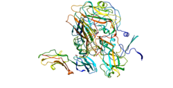
ICAM-1 appartient à la superfamille des immunoglobulines, qui comprend les anticorps, les récepteurs des cellules B (BCR) et les récepteurs des cellules T (TCR). ICAM-1 est une glycoprotéine transmembranaire possédant un seul domaine transmembranaire. Sa structure est caractérisée par une forte glycosylation et un domaine extracellulaire composé de nombreuses boucles créées par les ponts disulfure de la protéine. La structure secondaire dominante de la protéine est le feuillet bêta, ce qui conduit les chercheurs à émettre l'hypothèse de la présence de domaines de dimérisation au sein de l'ICAM-1.

Fonction

La protéine codée par ce gène est un type de molécule d'adhésion intercellulaire continu présente en faible concentration dans les membranes des leucocytes et les cellules endothéliales . Lors de la stimulation des cytokines, les concentrations augmentent considérablement. Lorsqu'il est activé, les leucocytes se lient aux cellules endothéliales via ICAM-1 / LFA-1, puis transmigrent dans les tissus.

Cible thérapeutique

Le lifitegrast est un antagoniste du LFA-1 empêchant son interaction avec l'ICAM-1 et en cours de test dans la sécheresse oculaire.

Interactions

Il a été montré qu'ICAM-1 interagit avec :

  • CD11a (en)[5],[6],[7] ;
  • EZR[8]
  • CD18[5],[9],[10]

Notes et références

  1. a b et c GRCh38: Ensembl release 89: ENSG00000090339 - Ensembl, May 2017
  2. a b et c GRCm38: Ensembl release 89: ENSMUSG00000037405 - Ensembl, May 2017
  3. « Publications PubMed pour l'Homme », sur National Center for Biotechnology Information, U.S. National Library of Medicine
  4. « Publications PubMed pour la Souris », sur National Center for Biotechnology Information, U.S. National Library of Medicine
  5. a et b Lu C, Takagi J, Springer TA, « Association of the membrane proximal regions of the alpha and beta subunit cytoplasmic domains constrains an integrin in the inactive state », The Journal of Biological Chemistry, vol. 276, no 18,‎ , p. 14642–48 (PMID 11279101, DOI 10.1074/jbc.M100600200)
  6. Shimaoka M, Xiao T, Liu JH, Yang Y, Dong Y, Jun CD, McCormack A, Zhang R, Joachimiak A, Takagi J, Wang JH, Springer TA, « Structures of the alpha L I domain and its complex with ICAM-1 reveal a shape-shifting pathway for integrin regulation », Cell, vol. 112, no 1,‎ , p. 99–111 (PMID 12526797, DOI 10.1016/S0092-8674(02)01257-6)
  7. Yusuf-Makagiansar H, Makagiansar IT, Hu Y, Siahaan TJ, « Synergistic inhibitory activity of alpha- and beta-LFA-1 peptides on LFA-1/ICAM-1 interaction », Peptides, vol. 22, no 12,‎ , p. 1955–62 (PMID 11786177, DOI 10.1016/S0196-9781(01)00546-0)
  8. Heiska L, Alfthan K, Grönholm M, Vilja P, Vaheri A, Carpén O, « Association of ezrin with intercellular adhesion molecule-1 and -2 (ICAM-1 and ICAM-2). Regulation by phosphatidylinositol 4, 5-bisphosphate », The Journal of Biological Chemistry, vol. 273, no 34,‎ , p. 21893–900 (PMID 9705328, DOI 10.1074/jbc.273.34.21893)
  9. Kotovuori A, Pessa-Morikawa T, Kotovuori P, Nortamo P, Gahmberg CG, « ICAM-2 and a peptide from its binding domain are efficient activators of leukocyte adhesion and integrin affinity », Journal of Immunology, vol. 162, no 11,‎ , p. 6613–20 (PMID 10352278)
  10. Huang C, Springer TA, « A binding interface on the I domain of lymphocyte function-associated antigen-1 (LFA-1) required for specific interaction with intercellular adhesion molecule 1 (ICAM-1) », The Journal of Biological Chemistry, vol. 270, no 32,‎ , p. 19008–16 (PMID 7642561, DOI 10.1074/jbc.270.32.19008)

Voir aussi

  • icône décorative Portail de la médecine
  • icône décorative Portail de la biologie cellulaire et moléculaire