PRKCQ
Protein-coding gene in the species Homo sapiens
| PRKCQ | |||||||||||||||||||||||||||||||||||||||||||||||||||
|---|---|---|---|---|---|---|---|---|---|---|---|---|---|---|---|---|---|---|---|---|---|---|---|---|---|---|---|---|---|---|---|---|---|---|---|---|---|---|---|---|---|---|---|---|---|---|---|---|---|---|---|
 | |||||||||||||||||||||||||||||||||||||||||||||||||||
| Identifiers | |||||||||||||||||||||||||||||||||||||||||||||||||||
| Aliases | PRKCQ, PRKCT, nPKC-theta, protein kinase C theta | ||||||||||||||||||||||||||||||||||||||||||||||||||
| External IDs | OMIM: 600448; MGI: 97601; HomoloGene: 21263; GeneCards: PRKCQ; OMA:PRKCQ - orthologs | ||||||||||||||||||||||||||||||||||||||||||||||||||
| |||||||||||||||||||||||||||||||||||||||||||||||||||
| |||||||||||||||||||||||||||||||||||||||||||||||||||
| |||||||||||||||||||||||||||||||||||||||||||||||||||
| |||||||||||||||||||||||||||||||||||||||||||||||||||
| |||||||||||||||||||||||||||||||||||||||||||||||||||
| Wikidata | |||||||||||||||||||||||||||||||||||||||||||||||||||
| |||||||||||||||||||||||||||||||||||||||||||||||||||


Protein kinase C theta (PKC-θ) is an enzyme that in humans is encoded by the PRKCQ gene.[5] PKC-θ, a member of serine/threonine kinases, is mainly expressed in hematopoietic cells[5] with high levels in platelets and T lymphocytes, where plays a role in signal transduction. Different subpopulations of T cells vary in their requirements of PKC-θ, therefore PKC-θ is considered as a potential target for inhibitors in the context of immunotherapy.[6]
Function
Protein kinase C (PKC) is a family of serine- and threonine-specific protein kinases that can be activated by the second messenger diacylglycerol. PKC family members phosphorylate a wide variety of protein targets and are known to be involved in diverse cellular signaling pathways. PKC family members also serve as major receptors for phorbol esters, a class of tumor promoters. Each member of the PKC family has a specific expression profile and is believed to play a distinct role. The protein encoded by this gene is one of the PKC family members. It is a calcium-independent and phospholipid-dependent protein kinase. This kinase is important for T-cell activation. It is required for the activation of the transcription factors NF-kappaB and AP-1, and may link the T cell receptor (TCR) signaling complex to the activation of the transcription factors.[7] PKC-θ also play a role in the apoptosis of lymphoid cells where it negatively influence and delay the aggregation of spectrin in an early phase of apoptosis.[8]
The role of PKC-θ in T cells
PKC-θ has a role in the transduction of signals in T cells, the kinase influences their activation, survival and growth. PKC-θ is important in the signal pathway integrating signals from TCR and CD28 receptors. A junction between an APC (an antigen presenting cell) and a T cell through their TCR and MHC receptors forms an immunological synapse. The active PKC-θ is localized in immunological synapse of T cells between the cSMAC (central supramolecular activation cluster containing TCR) and pSMAC (peripheral supramolecular activation cluster containing LFA-1 and ICAM-1). In regulatory T cells, PKC-θ is depleted from the region of immunological synapse, whereas in effector T cells, PKC-θ is present.[6] As a result of co-stimulation by CD28 and TCR, PKC-θ is sumoylated by SUMO1 predominantly on the sites Lys325 and Lys506. Sumoylation is important because of forming of the immunological synapse.[9] Subsequently, PKC-θ phosphorylates SPAK (STE20/SPS1-related, proline alanine-rich kinase) that activates the transcription factor AP-1 (activating protein-1). PKC-θ also initiates the assembly of proteins Carma-1, Bcl-10 and Malt-1 by phosphorylation of Carma-1. This complex of three proteins activates the transcription factor NF-κB (nuclear factor-κB). Furthermore, PKC-θ plays a role in the activation of transcription factor NF-AT (nuclear factor of activated T cells).[10] Thus, PKC-θ promotes inflammation in effector T cells.[6] PKC-θ plays a role in the activation of ILC2 and contribute to the proliferation of Th2 cells.[11] The kinase PKC-θ is crucial for function of Th2 and Th17.[6] Moreover, PKC-θ can translocate itself to the nucleus and by phosphorylation of histones increases the accessibility of transcriptional-memory-responsive genes in memory T cells.[12] PKC-θ plays a role in anti-tumor activity of NK cells. It was observed that in mice without PKC-θ, MHCI-deficient tumors are more often.[13]
The possible application of its inhibitors
Properties of PKC-θ make PKC-θ a good target for therapy in order to reduce harmful inflammation mediated by Th17 (mediating autoimmune diseases) or by Th2 (causing allergies)[11] without diminishing the ability of T cells to get rid of viral-infected cells. Inhibitors could be used in T-cell mediated adaptive immune responses. Inhibition of PKC-θ downregulates transcription factors (NF-κB, NF-AT) and cause lower production of IL-2. It was observed that animals without PKC-θ are resistant to some autoimmune diseases.[6] PKC-θ could be a target of inhibitors in the therapy of allergies.
The problem is that inhibitors of PKC-θ targeting catalytic sites may have toxic effects because of low specificity (catalytic sites among PKCs are very similar). Allosteric inhibitors have to be more specific to concrete isoforms of PKC.[6] s.
Interactions
PRKCQ has been shown to interact with:
PRKCQ has been shown to phosphorylate CARD11 as part of the NF-κB signaling pathway.[18]
Inhibitors
- (R)-2-((S)-4-(3-Chloro-5-fluoro-6-(1H-pyrazolo[3,4-b]pyridin- 3-yl)pyridin-2-yl)piperazin-2-yl)-3-methylbutan-2-ol[19]
See also
References
- ^ a b c GRCh38: Ensembl release 89: ENSG00000065675 – Ensembl, May 2017
- ^ a b c GRCm38: Ensembl release 89: ENSMUSG00000026778 – Ensembl, May 2017
- ^ "Human PubMed Reference:". National Center for Biotechnology Information, U.S. National Library of Medicine.
- ^ "Mouse PubMed Reference:". National Center for Biotechnology Information, U.S. National Library of Medicine.
- ^ a b Baier G, Telford D, Giampa L, Coggeshall KM, Baier-Bitterlich G, Isakov N, Altman A (April 1993). "Molecular cloning and characterization of PKC theta, a novel member of the protein kinase C (PKC) gene family expressed predominantly in hematopoietic cells". J Biol Chem. 268 (7): 4997–5004. doi:10.1016/S0021-9258(18)53494-3. PMID 8444877.
- ^ a b c d e f Zanin-Zhorov A, Dustin ML, Blazar BR (2011). "PKC-θ function at the immunological synapse: prospects for therapeutic targeting". Trends in Immunology. 32 (8): 358–363. doi:10.1016/j.it.2011.04.007. PMC 3573858. PMID 21733754.
- ^ "Entrez Gene: PRKCQ protein kinase C, theta".
- ^ Michalczyk I, Toporkiewicz M, Dubielecka PM, Chorzalska A, Sikorski AF (2016). "PKC-θ is a negative regulator of TRAIL-induced and FADD-mediated apoptotic spectrin aggregation". Folia Histochemica et Cytobiologica. 54 (1): 1–13. doi:10.5603/FHC.a2016.0006. PMID 27094638.
- ^ Wang XD, Gong Y, Chen ZL, Gong BN, Xie JJ, Zhong CQ, Wang QL, Diao LH, Xu A (2015). "TCR-induced sumoylation of the kinase PKC-θ controls T cell synapse organization and T cell activation". Nature Immunology. 16 (11): 1195–1203. doi:10.1038/ni.3259. ISSN 1529-2916. PMID 26390157. S2CID 21498259.
- ^ Zeng Q, Luo P, Gu J, Liang B, Liu Q, Zhang A (2017). "PKC θ-mediated Ca 2+ /NF-AT signalling pathway may be involved in T-cell immunosuppression in coal-burning arsenic-poisoned population". Environmental Toxicology and Pharmacology. 55: 44–50. doi:10.1016/j.etap.2017.08.005. PMID 28823652.
- ^ a b Madouri F, Chenuet P, Beuraud C, Fauconnier L, Marchiol T, Rouxel N, Ledru A, Gallerand M, Lombardi V (2017). "Protein kinase Cθ controls type 2 innate lymphoid cell and T H 2 responses to house dust mite allergen". Journal of Allergy and Clinical Immunology. 139 (5): 1650–1666. doi:10.1016/j.jaci.2016.08.044. PMID 27746240.
- ^ Li J, Hardy K, Phetsouphanh C, Tu WJ, Sutcliffe EL, McCuaig R, Sutton CR, Zafar A, Munier CM (2016-06-15). "Nuclear PKC-θ facilitates rapid transcriptional responses in human memory CD4+ T cells through p65 and H2B phosphorylation". J Cell Sci. 129 (12): 2448–2461. doi:10.1242/jcs.181248. ISSN 0021-9533. PMC 4920249. PMID 27149922.
- ^ Anel A, Aguiló JI, Catalán E, Garaude J, Rathore MG, Pardo J, Villalba M (2012). "Protein Kinase C-θ (PKC-θ) in Natural Killer Cell Function and Anti-Tumor Immunity". Frontiers in Immunology. 3: 187. doi:10.3389/fimmu.2012.00187. ISSN 1664-3224. PMC 3389606. PMID 22783260.
- ^ Bauer B, Krumböck N, Fresser F, Hochholdinger F, Spitaler M, Simm A, Uberall F, Schraven B, Baier G (August 2001). "Complex formation and cooperation of protein kinase C theta and Akt1/protein kinase B alpha in the NF-kappa B transactivation cascade in Jurkat T cells". J. Biol. Chem. 276 (34): 31627–34. doi:10.1074/jbc.M103098200. PMID 11410591.
- ^ Ron D, Napolitano EW, Voronova A, Vasquez NJ, Roberts DN, Calio BL, Caothien RH, Pettiford SM, Wellik S, Mandac JB, Kauvar LM (July 1999). "Direct interaction in T-cells between thetaPKC and the tyrosine kinase p59fyn". J. Biol. Chem. 274 (27): 19003–10. doi:10.1074/jbc.274.27.19003. PMID 10383400.
- ^ Witte S, Villalba M, Bi K, Liu Y, Isakov N, Altman A (January 2000). "Inhibition of the c-Jun N-terminal kinase/AP-1 and NF-kappaB pathways by PICOT, a novel protein kinase C-interacting protein with a thioredoxin homology domain". J. Biol. Chem. 275 (3): 1902–9. doi:10.1074/jbc.275.3.1902. PMID 10636891.
- ^ Hehner SP, Li-Weber M, Giaisi M, Dröge W, Krammer PH, Schmitz ML (April 2000). "Vav synergizes with protein kinase C theta to mediate IL-4 gene expression in response to CD28 costimulation in T cells". J. Immunol. 164 (7): 3829–36. doi:10.4049/jimmunol.164.7.3829. PMID 10725744.
- ^ Takeda K, Harada Y, Watanabe R, Inutake Y, Ogawa S, Onuki K, Kagaya S, Tanabe K, Kishimoto H, Abe R (December 2008). "CD28 stimulation triggers NF-kappaB activation through the CARMA1-PKCtheta-Grb2/Gads axis". Int. Immunol. 20 (12): 1507–15. doi:10.1093/intimm/dxn108. PMID 18829987.
- ^ Jimenez JM, Boyall D, Brenchley G, Collier PN, Davis CJ, Fraysse D, Keily SB, Henderson J, Miller A, Pierard F, Settimo L, Twin HC, Bolton CM, Curnock AP, Chiu P, Tanner AJ, Young S (2013). "Design and optimization of selective protein kinase C θ (PKCθ) inhibitors for the treatment of autoimmune diseases". J. Med. Chem. 56 (5): 1799–810. doi:10.1021/jm301465a. PMID 23398373.
Further reading
- Meller N, Altman A, Isakov N (1998). "New perspectives on PKCtheta, a member of the novel subfamily of protein kinase C." Stem Cells. 16 (3): 178–92. doi:10.1002/stem.160178. PMID 9617893. S2CID 83896240.
- Greenway AL, Holloway G, McPhee DA, Ellis P, Cornall A, Lidman M (2004). "HIV-1 Nef control of cell signalling molecules: multiple strategies to promote virus replication". J. Biosci. 28 (3): 323–35. doi:10.1007/BF02970151. PMID 12734410. S2CID 33749514.
- Ruegg CL, Strand M (1991). "A synthetic peptide with sequence identity to the transmembrane protein GP41 of HIV-1 inhibits distinct lymphocyte activation pathways dependent on protein kinase C and intracellular calcium influx". Cell. Immunol. 137 (1): 1–13. doi:10.1016/0008-8749(91)90051-C. PMID 1832084.
- Chowdhury IH, Koyanagi Y, Kobayashi S, Hamamoto Y, Yoshiyama H, Yoshida T, Yamamoto N (1990). "The phorbol ester TPA strongly inhibits HIV-1-induced syncytia formation but enhances virus production: possible involvement of protein kinase C pathway". Virology. 176 (1): 126–32. doi:10.1016/0042-6822(90)90237-L. PMID 1970444.
- Ruegg CL, Strand M (1990). "Inhibition of protein kinase C and anti-CD3-induced Ca2+ influx in Jurkat T cells by a synthetic peptide with sequence identity to HIV-1 gp41". J. Immunol. 144 (10): 3928–35. doi:10.4049/jimmunol.144.10.3928. PMID 2139676.
- Jakobovits A, Rosenthal A, Capon DJ (1990). "Trans-activation of HIV-1 LTR-directed gene expression by tat requires protein kinase C." EMBO J. 9 (4): 1165–70. doi:10.1002/j.1460-2075.1990.tb08223.x. PMC 551792. PMID 2182321.
- Fields AP, Bednarik DP, Hess A, May WS (1988). "Human immunodeficiency virus induces phosphorylation of its cell surface receptor". Nature. 333 (6170): 278–80. Bibcode:1988Natur.333..278F. doi:10.1038/333278a0. PMID 3259291. S2CID 4254146.
- Chirmule N, Goonewardena H, Pahwa S, Pasieka R, Kalyanaraman VS, Pahwa S (1995). "HIV-1 envelope glycoproteins induce activation of activated protein-1 in CD4+ T cells". J. Biol. Chem. 270 (33): 19364–9. doi:10.1074/jbc.270.33.19364. PMID 7642615.
- Chang JD, Xu Y, Raychowdhury MK, Ware JA (1993). "Molecular cloning and expression of a cDNA encoding a novel isoenzyme of protein kinase C (nPKC). A new member of the nPKC family expressed in skeletal muscle, megakaryoblastic cells, and platelets". J. Biol. Chem. 268 (19): 14208–14. doi:10.1016/S0021-9258(19)85228-6. PMID 7686153.
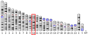
- Erdel M, Baier-Bitterlich G, Duba C, Isakov N, Altman A, Utermann G, Baier G (1995). "Mapping of the human protein kinase C-theta (PRKCQ) gene locus to the short arm of chromosome 10 (10p15) by FISH". Genomics. 25 (2): 595–7. doi:10.1016/0888-7543(95)80068-W. PMID 7790001.
- Ward NE, Gravitt KR, O'Brian CA (1995). "Inhibition of protein kinase C by a synthetic peptide corresponding to cytoplasmic domain residues 828-848 of the human immunodeficiency virus type 1 envelope glycoprotein". Cancer Lett. 88 (1): 37–40. doi:10.1016/0304-3835(94)03610-U. PMID 7850771.
- Gupta S, Aggarwal S, Kim C, Gollapudi S (1994). "Human immunodeficiency virus-1 recombinant gp120 induces changes in protein kinase C isozymes--a preliminary report". Int. J. Immunopharmacol. 16 (3): 197–204. doi:10.1016/0192-0561(94)90013-2. PMID 8206685.
- Parada NA, Cruikshank WW, Danis HL, Ryan TC, Center DM (1996). "IL-16- and other CD4 ligand-induced migration is dependent upon protein kinase C." Cell. Immunol. 168 (1): 100–6. doi:10.1006/cimm.1996.0054. PMID 8599832.
- Conant K, Ma M, Nath A, Major EO (1996). "Extracellular human immunodeficiency virus type 1 Tat protein is associated with an increase in both NF-kappa B binding and protein kinase C activity in primary human astrocytes". J. Virol. 70 (3): 1384–9. doi:10.1128/JVI.70.3.1384-1389.1996. PMC 189957. PMID 8627654.
- Smith BL, Krushelnycky BW, Mochly-Rosen D, Berg P (1996). "The HIV nef protein associates with protein kinase C theta". J. Biol. Chem. 271 (28): 16753–7. doi:10.1074/jbc.271.17.9906. PMID 8663223.
- Meller N, Liu YC, Collins TL, Bonnefoy-Bérard N, Baier G, Isakov N, Altman A (1996). "Direct interaction between protein kinase C theta (PKC theta) and 14-3-3 tau in T cells: 14-3-3 overexpression results in inhibition of PKC theta translocation and function". Mol. Cell. Biol. 16 (10): 5782–91. doi:10.1128/MCB.16.10.5782. PMC 231579. PMID 8816492.
- Holmes AM (1996). "In vitro phosphorylation of human immunodeficiency virus type 1 Tat protein by protein kinase C: evidence for the phosphorylation of amino acid residue serine-46". Arch. Biochem. Biophys. 335 (1): 8–12. doi:10.1006/abbi.1996.0476. PMID 8914829.
- Monks CR, Kupfer H, Tamir I, Barlow A, Kupfer A (1997). "Selective modulation of protein kinase C-theta during T-cell activation". Nature. 385 (6611): 83–6. Bibcode:1997Natur.385...83M. doi:10.1038/385083a0. PMID 8985252. S2CID 4255930.
- Datta R, Kojima H, Yoshida K, Kufe D (1997). "Caspase-3-mediated cleavage of protein kinase C theta in induction of apoptosis". J. Biol. Chem. 272 (33): 20317–20. doi:10.1074/jbc.272.33.20317. PMID 9252332.
- v
- t
- e
PDB gallery
-
 1xjd: Crystal Structure of PKC-theta complexed with Staurosporine at 2A resolution

Portal:
 Biology


