Layer of rods and cones
| Layer of rods and cones | |
|---|---|
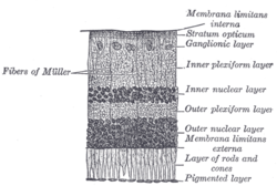 Section of retina. (Layer of rods and cones labeled at right, second from the bottom.) | |
 Plan of retinal neurons. (Layer of rods and cones labeled at left, at the bottom.) | |
| Details | |
| Identifiers | |
| Latin | Stratum photosensorium retinae |
| Anatomical terminology [edit on Wikidata] | |
The elements composing the layer of rods and cones (Jacob's membrane) in the retina of the eye are of two kinds, rod cells and cone cells, the former being much more numerous than the latter except in the macula lutea.
Jacob's membrane is named after Irish ophthalmologist Arthur Jacob, who was the first to describe this nervous layer of the retina. [1]
References
- ^ Somerville-Large, L. B. (1948). "Br J Ophthalmol: first published as 10.1136/bjo.32.9.601". The British Journal of Ophthalmology. 32 (9): 601–17. doi:10.1136/bjo.32.9.601. PMC 512141. PMID 18170498.
 This article incorporates text in the public domain from page 1017 of the 20th edition of Gray's Anatomy (1918)
External links
- Histology image: 07902loa – Histology Learning System at Boston University
- v
- t
- e
Anatomy of the globe of the human eye
(outer)
| Sclera |
|
|---|---|
| Cornea |
|

tunic (middle)
| Choroid | |
|---|---|
| Ciliary body | |
| Iris |
| Layers | |
|---|---|
| Cells |
|
| Other |
of the eye
| Anterior segment | |
|---|---|
| Posterior segment |
- Keratocytes
- Ocular immune system
- Optical coherence tomography
- Eye care professional
- Eye disease
- Refractive error
- Accommodation
- Physiological Optics
- Visual perception
 | This article about the eye is a stub. You can help Wikipedia by expanding it. |
- v
- t
- e
Portal:
 Anatomy


